Presentation
A 47-year-old male driver with a known 15-year history of diabetes mellitus and a 20-year smoking history (five pack-years) presented to the emergency treatment unit with a severe, generalized headache. The headache had lasted four hours, awakened him from sleep, and was most intense in the frontotemporal region. It was described as tightening in nature, continuous, and associated with nausea, two episodes of vomiting, and a sensation of faintness. He denied experiencing photophobia, phonophobia, chest discomfort, neck or arm pain, dyspnea, or sweating. At home, he had taken 1 g of paracetamol and applied Ayurvedic balm without symptom relief. Two days earlier, he had a mild fever and throat irritation for which he was prescribed paracetamol, diclofenac sodium, amoxicillin, and omeprazole by a private clinic. Those symptoms had subsided the day before presentation. His diabetes was being managed with Mixtard 30 insulin for the past four years following initial metformin therapy. However, his adherence was poor, and clinic follow-up was irregular. He often self-medicated and had not been screened for diabetic complications. His past surgical history included incision and drainage of abscesses on the left leg and right groin. Both parents had diabetes, and his father had died due to alcoholic cirrhosis.
Examination
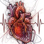
On admission, the patient was afebrile but hemodynamically unstable, with a pulse rate of 50 bpm and blood pressure of 88/50 mmHg. Oxygen saturation was 100% on room air. Cardiovascular and respiratory examinations were normal. Neurological examination revealed no abnormalities. However, the ECG showed ST-segment elevation in leads II, III, and aVF, with reciprocal ST depression in leads V2–V4.These results indicate acute inferior ST-elevation myocardial infarction (STEMI) and first-degree atrioventricular block.
Diagnosis
A diagnosis of acute inferior STEMI with associated bradycardia was made.It was supported by ECG findings and clinical presentation.
Management
The patient received loading doses of aspirin (300 mg), clopidogrel (300 mg), atorvastatin (80 mg), and was given atropine (1.2 mg in divided doses) and an isoprenaline infusion at 5 µg/min. About 30 minutes later, his headache began to subside. The patient was stable and oriented upon transfer to the cardiology unit. His pulse increased (84 bpm) and blood pressure improved (100/60 mmHg).The isoprenaline infusion was continued, and insulin therapy was optimized. Emergency coronary angiography found a total thrombotic occlusion of the mid-right coronary artery (RCA) and a long-segment stenosis (up to 80%) in the proximal to mid-left anterior descending artery (LAD).It confirmed the multivessel coronary artery disease. Both lesions were successfully stented. Following revascularization, the patient's headache resolved completely, and repeat ECG showed resolution of ST-elevation. Transthoracic echocardiography reported an ejection fraction of 55%.It also showed abnormal regional wall motion in the inferior wall. However, there was no pericardial effusion. Initial laboratory results showed markedly elevated serum troponin I of 6.14 ng/mL (normal <0.04 ng/mL).The patients had elevated serum creatinine (1.85 mg/dL and imrpoved to 1.1 mg/dL the next day).Liver transaminases were also raised on admission (AST: 128 U/L, ALT: 123 U/L) but declined within 48 hours (AST: 38 U/L, ALT: 65 U/L).Abdominal ultrasound findings were unremarkable.
Follow-Up
The patient was discharged three days post-admission on dual antiplatelet therapy (aspirin and clopidogrel), atorvastatin, bisoprolol, enalapril, and insulin (Mixtard 30).At his two-week and monthly follow-ups, the patient remained asymptomatic and returned to his routine activities with no new complaints.