Patient Overview
A 51-year-old man presented with a two-week history of left-sided limb weakness. Cranial magnetic resonance imaging (MRI) confirmed an acute lacunar infarction involving the right basal ganglia. During evaluation, newly diagnosed type 2 diabetes mellitus (T2DM) was identified based on glycated hemoglobin (HbA1c) of 7.2%, a 2-hour oral glucose tolerance test (OGTT) value ≥11.1 mmol/L, negative islet autoantibodies, and preserved fasting C-peptide levels. The patient was started on aspirin 100 mg once daily, atorvastatin calcium 20 mg nightly, and metformin 500 mg twice daily.
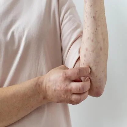
Ten days after treatment initiation, he developed progressively evolving cutaneous lesions over the trunk and extremities. The eruption initially appeared as scattered erythematous papules and subsequently evolved into round-to-oval targetoid lesions with central clearing, elevated borders, scaling, and mild pruritus. He denied fever, fatigue, arthralgia, lymphadenopathy, conjunctival involvement, oral ulcers, facial involvement, genital lesions, blistering, or erosions. The patient was admitted for evaluation of the rash.
He denied recent infection, travel history, exposure to new topical products, smoking, or alcohol use. His medical history was notable for a previous cerebral infarction, during which aspirin and atorvastatin had been used previously without adverse reactions before discontinuation two years earlier. He reported no known drug or food allergies, no history of asthma, urticaria, or allergic rhinitis, and no family history of genetic disorders.
Clinical Presentation
Physical examination demonstrated multiple scattered targetoid lesions with scaling predominantly distributed over the trunk and limbs, without mucosal, genital, or facial involvement. Neurological examination revealed left-sided limb weakness with muscle strength graded 4/5 and a positive left-sided pathological sign. Cardiovascular, respiratory, abdominal, and ophthalmologic examinations were unremarkable.
The absence of mucosal involvement, blistering, eosinophilia, or systemic organ dysfunction reduced the likelihood of Stevens–Johnson syndrome or Drug Reaction with Eosinophilia and Systemic Symptoms (DRESS) syndrome and supported a diagnosis consistent with drug-induced erythema multiforme.
Laboratory Findings
Laboratory evaluation demonstrated leukocytosis with a white blood cell count of 13.34 × 10⁹/L and neutrophils comprising 80.7% of leukocytes. C-reactive protein was elevated at 15.87 mg/L, and erythrocyte sedimentation rate measured 25 mm/h. Fasting plasma glucose was 9.69 mmol/L. Eosinophil percentage was 1.1%, with a normal absolute eosinophil count.
Additional diabetic evaluation demonstrated HbA1c of 7.2%, fasting C-peptide of 2.25 ng/mL, and negative glutamic acid decarboxylase antibody, insulin autoantibody, and islet cell antibody results, supporting the diagnosis of T2DM. Key laboratory findings are summarized in Table 1.
Table 1: Key Laboratory Findings
Parameter | Result | Reference Range |
White blood cell count | 13.34 × 10⁹/L ↑ | 3.97-9.15 × 10⁹/L |
Neutrophil percentage | 80.7% ↑ | 50.0%-70.0% |
Lymphocyte percentage | 12.4% ↓ | 20.0%-40.0% |
Eosinophil percentage | 1.1% ↓ | 0.02%-0.50% |
Erythrocyte sedimentation rate | 25 mm/h ↑ | 0-15 mm/h |
C-reactive protein | 15.87 mg/L ↑ | 0.00-3.00 mg/L |
Fasting plasma glucose | 9.69 mmol/L ↑ | 3.90-6.10 mmol/L |
2-hour OGTT glucose | 14.5 mmol/L ↑ | <7.8 mmol/L |
Glycated hemoglobin (HbA1c) | 7.2% ↑ | 4.2%-5.9% |
Fasting C-peptide | 2.25 ng/mL | 1.0-4.3 ng/mL |
Low-density lipoprotein cholesterol | 1.41 mmol/L | <1.4 mmol/L (target for patients with stroke) |
Creatine kinase | 215.0 IU/L ↑ | 25.0-195.0 IU/L |
Fibrinogen | 4.98 g/L ↑ | 2.00-4.00 g/L |
D-dimer | 1.05 μg/mL ↑ | 0.00-0.70 μg/mL |
Management
Given the absence of clinical evidence supporting connective tissue disease, malignancy, or infection, a drug-induced eruption was suspected, with metformin considered the most likely causative agent. All oral medications, including metformin, aspirin, and atorvastatin, were discontinued. Topical mometasone furoate gel and desonide ointment were applied to affected areas, and oral ebastine 20 mg once nightly was initiated for symptomatic relief.
Insulin therapy was initiated for glycemic management and consisted of insulin glulisine 4 U before meals combined with insulin degludec 10 U at bedtime. Insulin therapy was selected instead of alternative oral antihyperglycemic agents after discussion with the patient and based on his informed preference.
The rash and associated pruritus resolved completely within approximately two weeks after withdrawal of the oral medications. Following complete resolution of the eruption, aspirin and atorvastatin were sequentially reintroduced under close monitoring to maintain secondary prevention after cerebral infarction. No recurrence of cutaneous lesions occurred following rechallenge with either medication. The chronological sequence of clinical events, treatment modifications, and outcomes is summarized in Table 2.
Causality Assessment
Causality assessment using the Naranjo Adverse Drug Reaction Probability Scale yielded a score of 7, corresponding to a “probable” adverse drug reaction. Assessment using the World Health Organization–Uppsala Monitoring Centre (WHO–UMC) causality system was also categorized as “probable/likely.”
Table 2. Clinical Timeline
Timeline | Clinical Event |
Initial presentation | Left-sided limb weakness with MRI-confirmed acute lacunar infarction |
Baseline evaluation | Newly diagnosed T2DM based on HbA1c, OGTT, and preserved C-peptide |
Day 0 | Initiation of aspirin, atorvastatin, and metformin |
Day 10 | Development of targetoid cutaneous eruption with pruritus |
Hospital admission | Discontinuation of oral medications and initiation of topical therapy and insulin |
Approximately 2 weeks later | Complete resolution of rash and pruritus |
Post-recovery | Sequential rechallenge with aspirin and atorvastatin without recurrence |
Two-year follow-up | Stable glycemic control and no recurrent rash or ischemic events |
Follow-up
The patient was discharged in stable condition on insulin therapy, together with resumed aspirin and atorvastatin treatment.
During two years of follow-up, the patient remained free of recurrent skin lesions while continuing aspirin and atorvastatin therapy. Glycemic control remained stable, with fasting plasma glucose ranging from 5.5 to 6.8 mmol/L and HbA1c values ranging from 6.5% to 7.0%.
Follow-up laboratory monitoring demonstrated normal liver and kidney function, complete blood count, and C-reactive protein levels. No recurrent ischemic events were reported.