Patient Overview
A 78-year-old woman presented with progressively worsening involuntary movements involving the right upper limb for one month, followed by extension to the right lower limb. The movements persisted throughout the day, could not be voluntarily suppressed, and disappeared during sleep. These symptoms significantly affected her daily functioning. She denied fever, recent medication exposure, or family history of movement disorders, including Huntington’s disease. She also described polyuria and polydipsia over the preceding year.
Her medical history included hypertension and dyslipidemia, managed with amlodipine 10 mg once daily, telmisartan 80 mg once daily, and atorvastatin 40 mg nightly. She had not been previously diagnosed with diabetes mellitus.
Clinical Presentation
Physical examination identified choreiform movements confined to the right upper and lower limbs. Motor strength remained preserved at 5/5 in the affected extremities, with normal tone and reflexes. Sensory examination was normal. There were no signs suggestive of Parkinsonism, including rigidity or bradykinesia, and no evidence of cerebellar dysfunction. Cranial nerve and ocular examinations were also unremarkable.
Laboratory Findings
Laboratory evaluation demonstrated severe hyperglycemia without ketosis or metabolic acidosis. Mild hyponatremia and minimally elevated corrected calcium were noted, while renal and liver function parameters remained within normal limits. Key laboratory findings are summarized in Table 1.
Table 1. Key Laboratory Findings
Parameter | Result | Reference Range |
Random blood glucose | 22 mmol/L ↑ | – |
Glycated hemoglobin (HbA1c) | 15.9% ↑ | – |
Serum ketone | Negative | Negative |
Sodium | 134 mmol/L ↓ | 136-145 mmol/L |
Potassium | 3.8 mmol/L | 3.5-5.1 mmol/L |
Corrected calcium | 2.59 mmol/L ↑ | 2.20-2.55 mmol/L |
Phosphate | 0.89 mmol/L | 0.81-1.45 mmol/L |
Creatinine | 65 μmol/L | – |
eGFR | 78 mL/min/1.73 m² | – |
Alanine transaminase | 26.3 U/L | <35 U/L |
Arterial pH | 7.41 | – |
Serum bicarbonate | 27 mmol/L | 20-28 mmol/L |
C-reactive protein | 1.3 mg/L | <5 mg/L |
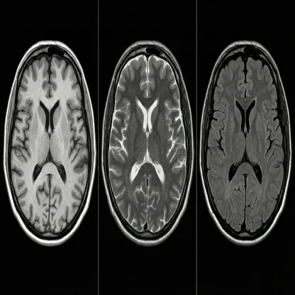
Neuroimaging Findings
Non-contrast computed tomography (CT) of the brain demonstrated hyperdensity involving the left caudate nucleus and left putamen. Magnetic resonance imaging (MRI) showed T1-weighted hyperintensity within the same regions, accompanied by low signal intensity on T2-weighted and T2 fluid-attenuated inversion recovery (FLAIR) sequences.
Gradient-recalled echo imaging demonstrated a blooming artifact without restricted diffusion on diffusion-weighted imaging or apparent diffusion coefficient mapping. Imaging also demonstrated volume loss involving the left caudate head and putamen, together with generalized cerebral and cerebellar atrophy. Small, scattered T2-weighted and FLAIR hyperintense foci were present within the subcortical frontoparietal white matter and periventricular regions, consistent with minimal small-vessel ischemic change (Fazekas grade 1).
The clinical findings, together with the characteristic imaging abnormalities, supported the diagnosis of diabetic striatopathy.
Management
The patient was started on intravenous insulin infusion for glycemic correction and was subsequently transitioned to subcutaneous insulin therapy. Haloperidol was initiated for symptomatic control of choreiform movements. During the one-week hospitalization, capillary blood glucose values remained between 8 and 10 mmol/L.
Improvement in glycemic status, together with symptomatic therapy, was associated with a reduction in the frequency and severity of the involuntary movements.
Follow-up
At the three-month neurology follow-up, choreiform movements were less frequent but had not completely resolved, requiring additional treatment with tetrabenazine. At six months, marked clinical improvement was observed, allowing discontinuation of tetrabenazine and gradual tapering of haloperidol.
Follow-up glycemic control improved substantially, with HbA1c decreasing to 4.9%.