Case Presentation
A 62-year-old man, status post coronary artery bypass grafting (CABG) one year earlier, arrived for pulmonary vein isolation (PVI) prompted by symptomatic paroxysmal atrial fibrillation (AF). Beyond CABG, his history stood clean, free of renal issues. His BMI registered at 26.9, with an entirely normal physical exam. Transthoracic echocardiography (TTE) confirmed preserved left ventricular ejection fraction and no left atrial dilation. Pre-procedure CT unveiled a prominent 37 mm left-sided common ostium alongside two right pulmonary veins (Figure 1). Procedure-day labs remained unremarkable (eGFR 80 mL/min/1.73 m², creatinine 89 µmol/L).
Procedure and Initial Course
The team deployed a 35 mm pentaspline Farapulse catheter during deep sedation via propofol and esketamine—chosen to match the oversized ostium seen on CT (Figure 1). Dual femoral vein access secured at 09:33 led straight to transseptal puncture at 09:35, followed by 0.5 mg atropine. Adhering to protocol, 40 applications (two 'olive' pulses per vein) secured full pulmonary vein isolation, verified by intracardiac echocardiography ensuring solid tissue contact. Sheaths exited at 10:05 amid activated clotting time exceeding 400 s. Vital signs held firm: starting blood pressure 109/75 mmHg dipped briefly to 95/73 mmHg before the initial pulse, then rebounded to 114/83 mmHg as sedation lifted. Recovery proved smooth, culminating in same-day discharge at 17:18.
Delayed Complication
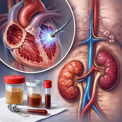
Hours later, cola-tinted urine emerged (Figure 2), spurring an emergency call at 22:30. The on-call resident couldn't connect initially but advised ample fluids (3 L/day) and close watch the next morning. By follow-up, urine cleared, yet output dwindled, malaise set in, and weight climbed 3 kg—signaling urgent return.
Examination and Diagnosis
Emergent labs exposed acute kidney injury (Table 1), marked by surging creatinine, modest lactate dehydrogenase (LDH) elevation, and intact haptoglobin (single assay). Renal ultrasound dismissed blockage, revealing typical kidney dimensions and echoes. Urinalysis spotted sparse pigmented granular casts. This profile pointed to post-PVI acute kidney injury, despite procedural haemodynamic steadiness, evoking pigment-driven renal insult.
Management
Initial therapy centered on 3 L intravenous fluids for support. As oliguria worsened, bumetanide 5 mg twice daily aimed to boost diuresis but halted at full anuria. Four haemodialysis runs followed, targeting 600–1400 mL ultrafiltration each (Figure 3).
Outcome and Discharge
Spontaneous urination restarted a week post-procedure, ushering a polyuric surge. Serial 24-hour collections tracked creatinine clearance rebound. Discharge came two weeks into admission, dialysis-free. Two months later, kidney function mirrored baseline (eGFR 66 mL/min/1.73 m², creatinine 104 µmol/L).
Featured
Off
Page Content
#ffffff
Anonymous user
On
Authenticated user
On
Premium
On
Paid / Sponsored
On
Key highlights
- Moderate pulsed field ablation (PFA) use can still harm the kidneys significantly during pulmonary vein isolation (PVI) under certain conditions.
- Large common ostia in the left atrium raise the risk of local red blood cell damage from electroporation, especially with poor catheter contact.
- Detect renal problems early, manage fluids aggressively, and consult nephrology quickly to aid kidney recovery.
Source
Mulder BA, Eisenga MF, Eijgelsheim M, Blaauw Y. Unexpected acute kidney injury requiring dialysis after routine pulsed field pulmonary vein isolation: a case report. Eur Heart J Case Rep. 2025 Dec 22;10(1):ytaf669. doi: https://doi.org/10.1093/ehjcr/ytaf669
Thumbnail
Schedule Date & Time
Speciality
Currency
Sub Speciality
Sub Sub Speciality
Short Description
A 62-year-old man with prior CABG underwent PVI for paroxysmal AF using Farapulse catheter. Despite normal pre-procedure renal function, he developed acute kidney injury with cola urine, requiring dialysis but full recovery.
User Segments
Release Date
Featured Order
0
Is Paid
0
Send Notification
Off