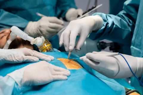
Percutaneous left heart interventions continue to expand across electrophysiology, structural heart disease, and heart failure care, increasing the use of transseptal puncture (TSP) in contemporary cardiovascular practice. Although TSP is widely performed in procedures such as atrial fibrillation ablation and transcatheter mitral interventions, it remains associated with potentially life-threatening complications.
The consensus statement provides practical recommendations for safe and efficient TSP, focusing on imaging-guided septal access, procedural technique, anticoagulation management, complication prevention, and management of complex anatomical and procedural scenarios.
Key Consensus Practice Recommendations
Advised To Do
- Follow recommendations according to the indication of the planned procedure when performing transseptal puncture (TSP).
- Use a transseptal approach for all endocardial left atrial (LA) interventional procedures.
- Perform cardiac imaging before TSP, including at minimum transthoracic echocardiography (TTE), to rule out major anatomical variants that may prevent safe and/or successful TSP.
- Perform atrial fibrillation (AF) ablation under uninterrupted oral anticoagulation (OAC) with vitamin K antagonists (VKAs) or uninterrupted/minimally interrupted direct oral anticoagulant (DOAC) therapy.
- Establish vascular access under ultrasound guidance to reduce vascular complications.
- Perform TSP with intraprocedural imaging guidance using fluoroscopy and/or transesophageal echocardiography (TEE) or intracardiac echocardiography (ICE).
- Confirm LA access during fluoroscopy-guided TSP using blood aspiration, pressure monitoring, dye injection, guidewire insertion, or a combination of these modalities.
- Administer heparin during AF ablation procedures to maintain an activated clotting time (ACT) of at least 300 seconds.
- Carefully check the size and length compatibility of all components of the transseptal assembly before the procedure.
- When fluoroscopic guidance alone is used during TSP, use at least one catheter as an anatomical landmark.
- Keep an alternative imaging modality, including TEE or ICE, readily available to guide TSP.
- Use TEE or ICE guidance when fluoroscopy alone is insufficient to identify the fossa ovalis or when TSP targets a specific septal location.
- Use TEE or ICE for TSP guidance when these imaging modalities are planned during the procedure.
- Perform immediate percutaneous drainage in cases of cardiac tamponade.
- Administer protamine to reverse heparin in patients with cardiac tamponade after the aspiration rate through percutaneous drainage decreases significantly.
- In cases of aortic puncture with sheath advancement, maintain a wire within the aorta to facilitate rapid sheath reinsertion if pericardial effusion occurs.
- Continuously flush the TSP sheath with heparinized saline to prevent thromboembolism.
- Flush the sheath before, during, and after catheter insertion and catheter retraction to prevent air embolism.
- In patients with an atrial septal closure device (ASCD), review device characteristics and use additional imaging with TEE or ICE alongside fluoroscopy for detailed characterization of cardiac anatomy.
May Be Appropriate To Do
- Consider heparin administration before TSP to prevent thromboembolism.
- Consider the use of a wire-based radiofrequency (RF) system under ultrasound visualization in patients with thick and/or aneurysmal septa to improve puncture ease and procedural safety.
- Consider a transseptal approach during left ventricular (LV) catheter ablation procedures to reduce vascular complications, aortic damage, and symptomatic or asymptomatic cerebral lesions.
- In cases of cardiac tamponade, direct autotransfusion of aspirated pericardial blood may be considered.
- In cases of air embolism, facilitate air expulsion from the coronaries using high-rate pacing and/or norepinephrine administration.
Areas of Uncertainty
- The safety of removing the pericardial drain in the electrophysiology (EP) laboratory after drainage of pericardial blood causing cardiac tamponade remains uncertain.