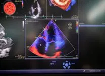
HFR MCE, which acquires images at more than 1000 frames per second, may enhance myocardial perfusion assessment by improving temporal resolution and reducing noise. An AHA 2025 Sessions study compared quantitative HFR MCE with conventional MCE for detecting OCAD and evaluating ischemia severity.
The study enrolled 25 eligible individuals, including 20 with high pre-test probability (PTP) and 5 with low PTP for OCAD. All underwent rest and vasodilator-stress imaging using both MCE techniques during intravenous microbubble infusion. Myocardial blood flow reserve (MBFR) was calculated from time–intensity curves, and MBFR <2.0 indicated reduced flow. Participants with high PTP also underwent invasive coronary angiography.
Coronary angiography confirmed OCAD in 27 vascular territories. HFR MCE identified reduced MBFR in 25 of these territories (93%), compared with 22 territories (81%) using conventional MCE. Among 12 normal vascular territories in low-PTP individuals, HFR MCE classified 7 as normal and conventional MCE classified 5 as normal. Overall accuracy for OCAD detection was 83% for HFR MCE and 69% for conventional imaging.
HFR MCE also identified more segments with reduced MBFR in individuals with OCAD and demonstrated a significantly lower hyperaemic myocardial blood flow value. These findings indicate that HFR MCE provides a more detailed assessment of ischemia extent and intensity while maintaining diagnostic accuracy comparable to conventional MCE.