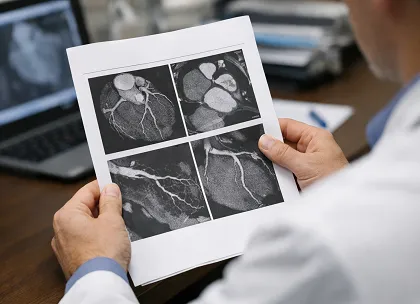
AI-guided quantitative coronary CT angiography (AI-QCT) showed higher discrimination for major adverse cardiovascular events (MACE) compared with clinical risk models in a multicenter observational cohort analysis published in JACC: Advances. Quantitative plaque assessment using coronary CT angiography (CCTA) has demonstrated strong correlation with intravascular imaging and prognostic relevance in coronary artery disease (CAD).
This multicenter, international cohort study from the CONFIRM2 registry included 3,551 patients (mean age 58.8 ± 12.5 years; 50.5% male) who underwent clinically indicated CCTA and were followed for a median of 4.27 years (interquartile range 3.47-5.08). Patients without cardiac symptoms or with prior CAD were excluded. AI-QCT analysis generated 24 variables across patient, vessel, and plaque levels, including luminal stenosis, remodeling index, and plaque volumes. The primary endpoint was MACE, defined as all-cause death, myocardial infarction (MI), stroke, congestive heart failure, late revascularization, and hospitalization for unstable angina. The secondary endpoint included all-cause death and MI.
During follow-up, 167 events (4.7%) were recorded. After excluding collinear variables, diameter stenosis (hazard ratio [HR] 1.25; 95% confidence interval [CI]: 1.18-1.32 per 10% increase) and noncalcified plaque volume (HR 1.07; 95% CI: 1.03-1.11 per 50 mm³ increase) were independently associated with MACE. In multivariable models, addition of AI-QCT measures increased area under the curve (AUC) from 0.63 (95% CI: 0.58-0.67) to 0.76 (95% CI: 0.77-0.80), P<0.001. Similar improvement was observed when AI-QCT was added to traditional risk factors, age, and sex (AUC 0.67 to 0.77; P<0.001). Discrimination also increased compared with the atherosclerotic cardiovascular disease risk score from 0.63 (95% CI: 0.58–0.68) to 0.75 (95% CI: 0.69-0.80; P<0.001). Comparable findings were reported for the secondary endpoint of all-cause death and MI.
AI-QCT-derived parameters were associated with higher discrimination of MACE compared with clinical models. Diameter stenosis and noncalcified plaque volume were identified as independent predictors of events.