Classic AFD results in more extensive cardiac involvement than the late-onset phenotype, according to a multimodality imaging study published in the International Journal of Cardiology. The investigation identified significant electrophysiologic, structural, and functional differences between the two groups.
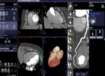
A total of 80 patients (40 classic, 40 late-onset) underwent echocardiography, electrocardiography (ECG), and cardiac magnetic resonance imaging (CMR). Age and sex balance were achieved using inverse probability of treatment weighting (IPTW). Classic AFD was associated with higher prevalence of delta waves, short PQ interval, pathological repolarization, elevated Sokolow–Lyon voltage criteria, and reduced T-wave amplitudes.
Cardiac imaging measures also reflected more advanced disease in the classic phenotype, including greater left ventricular diastolic dysfunction, more impaired left ventricular global longitudinal strain (LV GLS), larger left atrial (LA) volumes, and lower native T1 values. Classic AFD males demonstrated significantly worse LV GLS, LA strain, and higher LACI, while classic females presented with more pronounced LV diastolic dysfunction without marked differences in deformation indices.
Age-interaction modeling indicated faster deterioration of LACI with aging in classic AFD. Restricted cubic spline (RCS) analysis showed nonlinear associations between age and both left ventricular mass index (LVMI) and LV GLS in each phenotype, with slower progression beyond the early 40s.
These findings support more frequent and comprehensive cardiac assessment in individuals with classic AFD to identify and manage progression at earlier stages.