Early identification of retinal dysfunction is essential for preventing vision loss in adults with diabetes. A study published in Diabetology evaluated whether color perimetry can detect functional retinal changes before the onset of retinopathy.
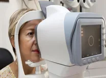
The study enrolled 40 age-matched adults grouped by (HbA1c: controls (≤5.6%), prediabetes (5.7-6.4%), and diabetes (≥6.5%). Additional variables included body mass index (BMI), body fat percentage, lipid levels, blood glucose, and blood pressure. Color perimetry measured chromatic thresholds at four retinal locations positioned three degrees from the fovea. Five conditions were tested: achromatic, red, green, blue, and yellow. Mars contrast sensitivity testing and L’Anthony D15 testing were also completed. Lens photographs excluded adults with cataracts.
HbA1c differed significantly among groups (p < 0.001). BMI and body fat percentage also differed (p = 0.046 and p = 0.020). Color perimetry detected group differences only in the yellow condition (p = 0.013). Achromatic thresholds correlated strongly with Mars contrast sensitivity (p < 0.001). HbA1c correlated with parvocellular function, measured by the red and green average (p = 0.014), and koniocellular function, measured by the blue and yellow average (p = 0.022).
These findings show that yellow chromatic thresholds may serve as an early functional marker of retinal changes in adults with diabetes before retinopathy is visible. Larger studies are needed to confirm the clinical utility of color perimetry.