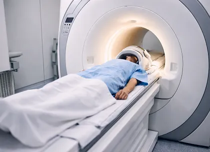
Cardiac magnetic resonance imaging (MRI) commonly relies on repeated breath-holding, which may be difficult for some patients and can prolong scan duration. A prospective study published in Radiology: Cardiothoracic Imaging evaluated whether a comprehensive free-breathing cardiac MRI protocol could improve efficiency while maintaining diagnostic performance compared with conventional breath-holding MRI.
The analysis included 605 participants (mean age 48 ± 17 years; 422 male) who underwent both free-breathing and breath-holding MRI acquisitions on a 3.0-T scanner between June 2024 and August 2024. The study assessed cine imaging, T1 and T2 mapping, flow imaging, and late gadolinium enhancement (LGE). Outcomes included total scan time, image quality, left ventricular (LV) functional measures, and tissue characterization.
The free-breathing protocol significantly reduced total scan duration compared with breath-holding MRI (20.9 ± 3.1 minutes vs 34.4 ± 7.5 minutes; P < .001). Cine, T1 mapping, and LGE acquisitions were 54%, 28%, and 33% faster, respectively. Image quality remained diagnostic across sequences. Cine scores were slightly lower, T1/T2 mapping and flow scores were similar, and LGE scores were higher with free-breathing MRI.
Interobserver agreement was strong (κ = 0.80-0.93). LV end-diastolic and end-systolic volumes were slightly higher, while LV ejection fraction was slightly lower with free-breathing MRI. However, agreement between methods remained excellent, with an intraclass correlation coefficient (ICC) of 0.98–0.99. Native T1 values and LGE mass were also slightly higher but closely aligned with breath-holding measurements.
The findings suggest that a comprehensive free-breathing cardiac MRI protocol may streamline imaging workflows while preserving diagnostic reliability across major cardiac MRI sequences.