Non-contrast imaging approaches for pulmonary perfusion assessment are being explored in suspected chronic thromboembolic pulmonary hypertension (CTEPH). This prospective, institutional review board (IRB)-approved study published in the Journal of Thoracic Imaging compared X-ray pulsatility index (XPI) with digital subtraction angiography (DSA) for pulmonary perfusion mapping.
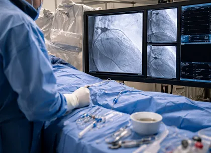
A total of 13 patients (6 male, 7 female) were enrolled between April 2023 and May 2024, including 18 lungs for analysis. Fluoroscopic imaging was performed at 70 kV and 30 frames per second during an 8-second breath-hold. Temporal pixel-level signals were analyzed using spectral decomposition, isolating signals at the heart rate to generate XPI-based perfusion maps.
Immediately after XPI acquisition, contrast-enhanced DSA was performed in the same projection. Perfusion maps from both modalities were segmented using a semi-automated method, and agreement was assessed using the Dice similarity score. All patients successfully completed the breath-hold protocol. Comparative analysis showed a mean Dice score of 0.70, indicating agreement between XPI and DSA in depicting regions of blood flow and perfusion.
XPI provided non-contrast perfusion mapping comparable to angiographic findings and may be used for pulmonary perfusion evaluation.