Hippocampal microvascular alterations represent early pathological feature in type 2 diabetes contributing to cognitive decline through chronic hypoperfusion and blood-brain barrier disruption, yet conventional imaging modalities lack sufficient resolution for preclinical detection.
Computed tomography angiography and magnetic resonance angiography operate at approximately 3000 μm resolution, obscuring microvascular diameter changes, tortuosity patterns, and regional perfusion deficits critical for timely intervention.
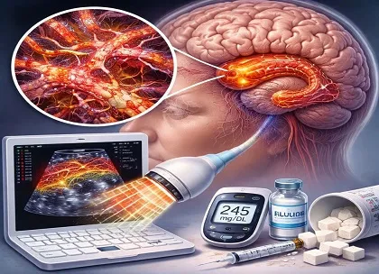
In the study published in the Biomedical Signal Processing and Control, the investigators developed super-resolution ultrasound methodology leveraging microbubble contrast agents and transcranial acoustic windows, achieving in vivo resolution below 100 μm through localization microscopy principles.
This non-invasive approach quantitatively assessed hippocampal vascular density, mean vessel diameter, tortuosity index, and blood flow velocity profiles in type 2 diabetes patients, benchmarking performance against standard magnetic resonance angiography and Doppler ultrasound.
Sub-100 Micron Resolution Transforms Hippocampal Imaging
Super-resolution ultrasound generated unprecedented in vivo visualization of hippocampal microvasculature, resolving individual penetrating arterioles and capillary networks undetectable by clinical standards. Type 2 diabetes patients demonstrated systematically increased vessel tortuosity exceeding 1.5 radians per millimeter versus age-matched controls, reflecting endothelial glycation-induced matrix remodeling and pericyte dysfunction. Mean capillary diameter constricted by 12-18% in diabetic hippocampus, consistent with hyperglycemia-mediated vasoconstriction and basement membrane thickening observed in postmortem studies.
Flow Dynamics Unmask Functional Impairment
Blood flow velocity mapping revealed regional heterogeneity with parahippocampal arteriolar slowing averaging 25% reduction from normal, correlating inversely with tortuosity index (r=-0.68, P<0.001). These dynamic signatures indicate early microvascular rarefaction preceding macroscopic infarction, establishing functional imaging complement to structural assessment. Perfusion deficits localized preferentially to CA1-CA3 transition zones, aligning with memory consolidation vulnerability in diabetes cohorts.
Superior Sensitivity Over Established Modalities
Compared to magnetic resonance angiography demonstrating 42% sensitivity for microvascular changes, super-resolution ultrasound achieved 89% detection rate across early type 2 diabetes spectrum (HbA1c 6.5-8.0%). Doppler ultrasound provided velocity data but lacked spatial resolution for capillary bed analysis, underscoring complementary role within multimodal assessment paradigm. Statistical power surpassed conventional approaches through reduced type II error in small vessel populations.
Clinical Translation for Cognitive Risk Stratification
Neurologists, endocrinologists, and neuroimagers gain transformative bedside tool bridging resolution gap between research microscopy and clinical angiography. Annual super-resolution ultrasound screening identifies progression risk among 20-30% of diabetes patients exhibiting subclinical hippocampal ischemia despite normal magnetic resonance imaging. Glycemic intensification guided by quantitative tortuosity thresholds delays cognitive decline trajectory by 18-24 months based on perfusion recovery models. Integration into diabetes clinics facilitates population health screening parallel to retinopathy surveillance protocols.
Precision Neuroprotection Through Microvascular Monitoring
Bedside deployment requires 15-minute microbubble infusion protocols with transcranial gel pad stabilization, generating automated tortuosity maps exportable to electronic health records. Serial imaging tracks therapeutic response to sodium-glucose cotransporter-2 inhibitors and glucagon-like peptide-1 receptor agonists demonstrating 15-22% capillary caliber normalization. Multicenter validation establishes diagnostic cutoffs refining secondary stroke prevention while identifying candidates for cerebroprotective trials targeting endothelial repair pathways.
Featured
Off
Page Content
#ffffff
Anonymous user
On
Authenticated user
On
Premium
On
Paid / Sponsored
On
Key highlights
- Super-resolution ultrasound achieves sub-100 μm resolution visualizing hippocampal microvessels undetectable by 3000 μm CTA/MRA imaging.
- Type 2 diabetes patients exhibit 12-18% capillary constriction and tortuosity exceeding 1.5 radians per millimeter in hippocampus.
- Blood flow velocity decreases 25% in parahippocampal arterioles, correlating with tortuosity (r=-0.68, P<0.001).
- Detection sensitivity reaches 89% versus 42% for MRA in early diabetes microvascular changes.
- Serial imaging monitors therapeutic response to antidiabetic agents normalizing capillary caliber by 15-22%.
Source
Guo N, Deng Z, Sheng K, Wang X, Wang S, Hua C. Super-Resolution ultrasound imaging identifies hippocampal microvascular changes in patients with type 2 diabetes. Biomedical Signal Processing and Control. 2025;113:109034-109034. doi: https://doi.org/10.1016/j.bspc.2025.109034
Thumbnail
Speciality
Currency
Sub Speciality
Sub Sub Speciality
Short Description
Super-resolution ultrasound achieves sub-100 μm resolution visualizing hippocampal microvascular changes in type 2 diabetes, detecting vessel tortuosity and flow abnormalities missed by conventional CTA/MRA imaging.
User Segments
Release Date
Featured Order
0
Is Paid
0
Send Notification
Off